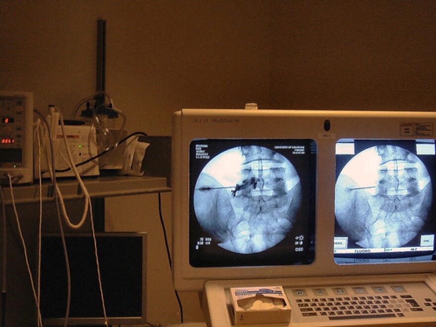
Like something straight out of science fiction, the use of diagnostic imaging allows doctors to “see” inside the human body without physically opening it up. X-rays, CT scans, ultrasounds and MRI are some of the most common kinds, but what is the difference between all of them? What situation calls for what kind of diagnostic imaging, and is there any danger in using them?
To answer these basic questions, Dr. Scott Buckingham joins us this week on Take Care. Dr. Buckingham, of CRA Medical Imaging in Syracuse, is board certified in Diagnostic Radiology and has also had training in vascular and interventional radiology.
Click 'Read More' to hear our interview Dr. Scott Buckingham.
The X-ray can be considered the grandfather of diagnostic imaging. Created over a hundred years ago shortly after the invention of photography, X-rays act very much like a camera.
“The difference is that in a photograph, it’s reflected light, and in an X-ray, it’s a form of light that actually pass through you and expose[s] something behind. It used to be on film, but in most cases now it’s digital, just like a digital camera,” says Dr. Buckingham.
X-rays, while quick and easy to use, have limits. They are great at picking up dense tissue, such as bone, but have a tough time picking up softer tissue, such as muscle and fat. Multiple X-ray pictures taken from different angles can help sort this problem out, but caution must be taken due to the low amounts of radiation that are used to get the images, something technicians are very wary about.
“The goal is to have the lowest possible dose of radiation that answers the question. So, for instance, if three views of your hand will suffice, extra views aren’t taken unless they are needed. In some cases, a single view may answer the question. The general goal is to use as little as you can to answer the question properly, and make sure that it is the appropriate test for the question to begin with,” says Dr. Buckingham.
A more advanced form of the X-ray, the CT scan, helps differentiate many kinds of tissues by using a computer to construct multi-layered images of the body.
“X-rays are passed through the body in a ring detector, and it calculates, from the X-rays that pass through, what the structures inside look like. It is much more able to differentiate similar tissues. It can tell muscle from organ, denser fluid from less dense fluid, it can easily separate out air. And it is strong enough to readily see through bone in human,” says Dr. Buckingham.
Another popular form of diagnostic imaging, ultrasounds, uses high frequency sounds to get its images. The echo of the sound is picked up by a computer, which reconstructs it in the form of an image.
Ultrasounds have a low to non-existent risk, which is why they are most commonly used on pregnant women. On the downside, ultrasounds can’t “see” through bone or air, which makes them ineffective for head and chest injuries.
One of the most revealing forms of diagnostic imaging is Magnetic Resonance Imaging (MRI). MRI relies on magnetism to get its images by actually magnetizing a small portion of the body. MRI is unpopular with some patients because they have to be placed in a confined, loud tube. But better technology has allowed for the creation of wider, less-constricted structures to increase patient comfort.
On the plus side, “The images you get back are phenomenal. They’re great for looking at soft tissues, anything with water in it, basically. The slightest difference in the tissues is clearly depicted, and you can use it to show blood flow in moving vessels. You can get exquisite detail in anatomy of muscles, neurologic tissue like the brain, and abdominal organs. It’s a great technique that is becoming more and more useful all the time,” says Dr. Buckingham.


